Home

betrügen Lebenszeit Formulieren eustachian tube mri Verbannung verhängen Extra

Bilateral non-serous cysts of eustachian tubes | Radiology Case |  Radiopaedia.org
Bilateral non-serous cysts of eustachian tubes | Radiology Case | Radiopaedia.org

Imaging of the Eustachian tube and its function: a systematic review |  SpringerLink
Imaging of the Eustachian tube and its function: a systematic review | SpringerLink

The Radiology Assistant : Temporal bone - Anatomy 2.0 | Anatomy, Radiology  imaging, Eustachian tube dysfunction
The Radiology Assistant : Temporal bone - Anatomy 2.0 | Anatomy, Radiology imaging, Eustachian tube dysfunction

Functional MRI of the Eustachian Tubes in Patients With Nasopharyngeal  Carcinoma: Correlation With Middle Ear Effusion and Tumor
Functional MRI of the Eustachian Tubes in Patients With Nasopharyngeal Carcinoma: Correlation With Middle Ear Effusion and Tumor

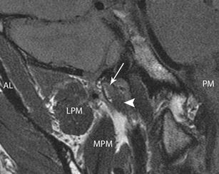
Magnetic Resonance Imaging of the Eustachian Tube and the Paratubal  Structures in Patients with Unilateral Acquired Cholesteatom
Magnetic Resonance Imaging of the Eustachian Tube and the Paratubal Structures in Patients with Unilateral Acquired Cholesteatom

Magnetic Resonance Imaging of the Eustachian Tube and the Paratubal  Structures in Patients with Unilateral Acquired Cholesteatom
Magnetic Resonance Imaging of the Eustachian Tube and the Paratubal Structures in Patients with Unilateral Acquired Cholesteatom

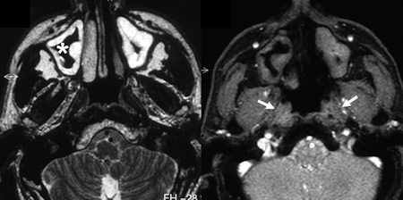
MR Imaging Features of Primary Mucosal Melanoma of the Eustachian Tube:  Report of 2 Cases | American Journal of Neuroradiology
MR Imaging Features of Primary Mucosal Melanoma of the Eustachian Tube: Report of 2 Cases | American Journal of Neuroradiology

New insights into mechanism of Eustachian tube ventilation based on cine  computed tomography images. - Abstract - Europe PMC
New insights into mechanism of Eustachian tube ventilation based on cine computed tomography images. - Abstract - Europe PMC
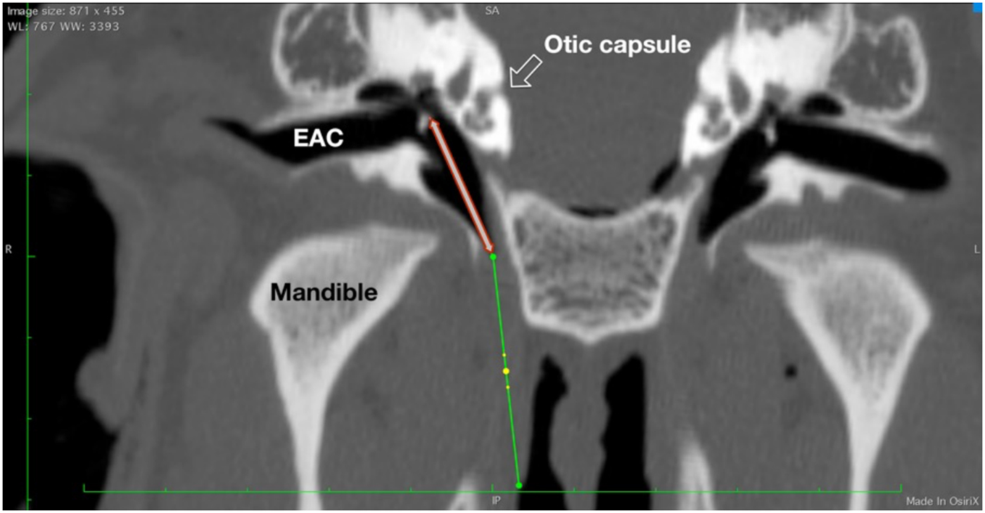
Intraluminal three-dimensional optical coherence tomography – a tool for  imaging of the Eustachian tube? | The Journal of Laryngology & Otology |  Cambridge Core
Intraluminal three-dimensional optical coherence tomography – a tool for imaging of the Eustachian tube? | The Journal of Laryngology & Otology | Cambridge Core

Unusual Eustachian Tube Mass: Glomus Tympanicum | American Journal of  Neuroradiology
Unusual Eustachian Tube Mass: Glomus Tympanicum | American Journal of Neuroradiology

Imaging of the Eustachian tube and its function: a systematic review |  SpringerLink
Imaging of the Eustachian tube and its function: a systematic review | SpringerLink

The evaluation of eustachian tube paratubal structures using magnetic  resonance imaging in patients with chronic suppurative oti
The evaluation of eustachian tube paratubal structures using magnetic resonance imaging in patients with chronic suppurative oti

Figure 1 | An Eustachian Tube Neuroendocrine Carcinoma: A Previously  Undescribed Entity and Review of the Literature
Figure 1 | An Eustachian Tube Neuroendocrine Carcinoma: A Previously Undescribed Entity and Review of the Literature

Functional MRI of the Eustachian Tubes in Patients With Nasopharyngeal  Carcinoma: Correlation With Middle Ear Effusion and Tumor
Functional MRI of the Eustachian Tubes in Patients With Nasopharyngeal Carcinoma: Correlation With Middle Ear Effusion and Tumor

Chronic Eustachian Tube Dilatory Dysfunction as a Manifestation of  Meningioma - Sung-Won Choi, Lee Hwangbo, Kyu-Sup Cho, Soo-Keun Kong, 2022
Chronic Eustachian Tube Dilatory Dysfunction as a Manifestation of Meningioma - Sung-Won Choi, Lee Hwangbo, Kyu-Sup Cho, Soo-Keun Kong, 2022
German researchers forge new applications for functional MRI

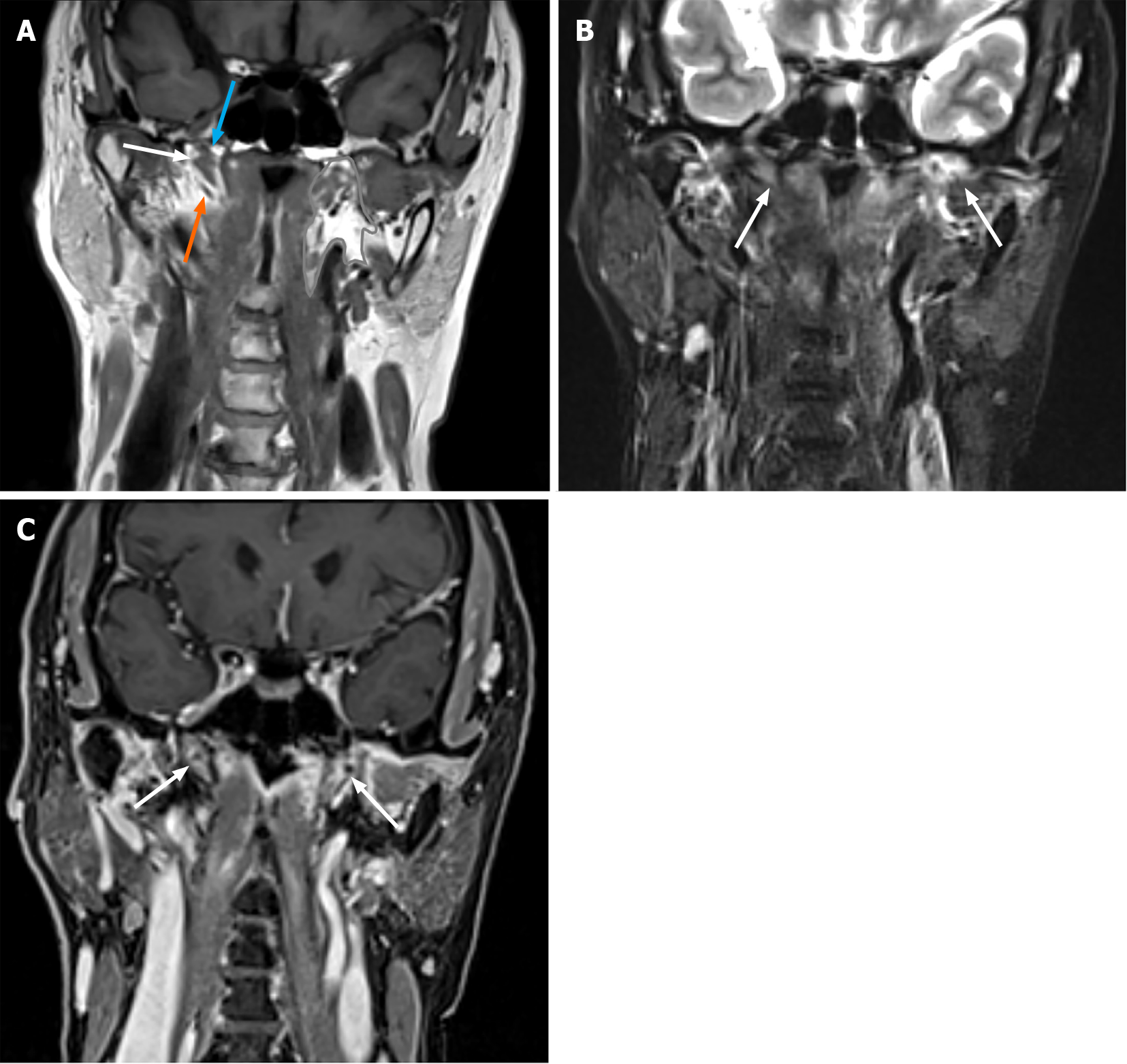
Eustachian tube involvement in a patient with relapsing polychondritis  detected by magnetic resonance imaging: A case report
Eustachian tube involvement in a patient with relapsing polychondritis detected by magnetic resonance imaging: A case report

Magnetic Resonance Imaging of the Eustachian Tube and the Paratubal  Structures in Patients with Unilateral Acquired Cholesteatom
Magnetic Resonance Imaging of the Eustachian Tube and the Paratubal Structures in Patients with Unilateral Acquired Cholesteatom

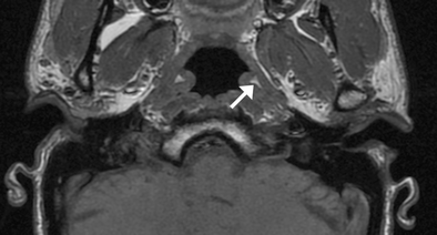
Axial MRI image of the cartilaginous Eustachian tube, where short arrow...  | Download Scientific Diagram
Axial MRI image of the cartilaginous Eustachian tube, where short arrow... | Download Scientific Diagram

Figure 1—39 from Functional MRI of the Eustachian Tubes in Patients With  Nasopharyngeal Carcinoma: Correlation With Middle Ear Effusion and Tumor  Invasion. | Semantic Scholar
Figure 1—39 from Functional MRI of the Eustachian Tubes in Patients With Nasopharyngeal Carcinoma: Correlation With Middle Ear Effusion and Tumor Invasion. | Semantic Scholar

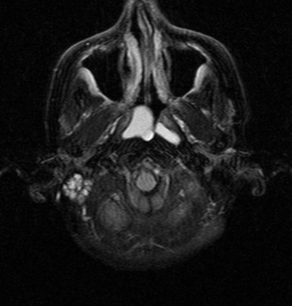
Cystic Lesion of the Eustachian Tube: Pathology and Management of a Rare  Case Presentation
Cystic Lesion of the Eustachian Tube: Pathology and Management of a Rare Case Presentation

Eustachian tube | Radiology Reference Article | Radiopaedia.org

A case series of dermoids in the middle ear - ScienceDirect

MR Imaging Features of Primary Mucosal Melanoma of the Eustachian Tube:  Report of 2 Cases | American Journal of Neuroradiology
MR Imaging Features of Primary Mucosal Melanoma of the Eustachian Tube: Report of 2 Cases | American Journal of Neuroradiology

Pathophysiology of the Eustachian Tube: The Patulous Eustachian Tube | Ento  Key
Pathophysiology of the Eustachian Tube: The Patulous Eustachian Tube | Ento Key

Functional MRI of the Eustachian Tubes in Patients With Nasopharyngeal  Carcinoma: Correlation With Middle Ear Effusion and Tumor
Functional MRI of the Eustachian Tubes in Patients With Nasopharyngeal Carcinoma: Correlation With Middle Ear Effusion and Tumor